| Call | (011) 4162-4583 | to schedule an eye exam |
Where the focus is on you
| Call | (011) 4162-4583 | to schedule an eye exam |
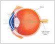
Diabetes is a disease, which affects the small blood vessels of all the organs of the body. As the disease progresses it inevitably involves the microvascular architecture of the body including the blood supply of the retina (sensory part of the eye). This manifestation of diabetes is called as diabetic retinopathy.
To simplify - It is a disease of the blood vessels of the retina due to diabetes.
People with diabetes have a higher risk of blindness and other various eye problems. Diabetes can effect the eyes and vision in a number of ways. It may lead to frequent fluctuations in vision, cataract in young age, decreased vision due to involvement of optic nerve, temporary paralysis of the muscles controlling the movement of eyes and thus double vision, incidence of glaucoma is higher in diabetics. The most significant complication of diabetes in eye is diabetic retinopathy and its complications.
Non-proliferative or background diabetic retinopathy: When blood vessels in the retina are damaged, they can leak fluid or bleed. This causes the retina to swell and form deposits called exudates. This is an early form of diabetic retinopathy and may not lead to any decrease in vision, but it can lead to other more serious forms of retinopathy that affect the vision.
Macular edema: The fluid and exudates collects in the macula (the part of the retina that allows us to see fine details), thus decreasing the vision. Sometimes there may be a macular edema without any loss of vision. Therefore it is important to have periodic checkup to detect and treat these conditions at an early stage.
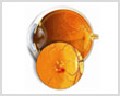
Bleeding from the retinal blood vessels due to diabetes
Proliferative Diabetic Retinopathy: This is an advanced stage of diabetic retinopathy, where the blood supply of retina is compromised. In response to this, new fragile blood vessels grow on the surface of the retina (neovascularization). These new vessels are very fragile and bleed easily. These may lead to serious vision problems if they bleed into the vitreous (the clear, jelly-like substance that fills the center of the eye) which is known as vitreous hemorrhage. This prevents the light from reaching the retina and thus can blur the vision.
The new blood vessels and the bleed into the vitreous can also cause scar tissue to develop, which can pull the retina away from the back of the eye. This is known as retinal detachment, and can lead to blindness if untreated.
In addition, abnormal blood vessels can grow on the iris (the colored part in the front of your eye, which can lead to glaucoma).
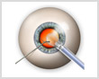
Huge strides have been made in the treatment of diabetic retinopathy. Treatments such as scatter photocoagulation, focal photocoagulation, and vitrectomy prevent blindness in most people. The sooner retinopathy is diagnosed; the more likely these treatments will be successful. The best results occur when sight is still normal.
In photocoagulation, the eye care professional makes tiny burns on the retina with a special laser. These burns seal the blood vessels and stop them from growing and leaking.
In scatter photocoagulation (also called panretinal photocoagulation); the eye care professional makes hundreds of burns in a polka-dot pattern on two or more occasions. Scatter photocoagulation reduces the risk of blindness from vitreous hemorrhage or detachment of the retina -- but it only works before bleeding or detachment has progressed very far. This treatment is also used for some kinds of glaucoma.
Side effects of scatter photocoagulation are usually minor. They include several days of blurred vision after each treatment and possible loss of side (peripheral) vision.
In focal photocoagulation, the eye care professional aims the laser precisely at leaking blood vessels in the macula. This procedure does not cure blurry vision caused by macular edema. But it does keep it from getting worse.
When the retina has already detached or a lot of blood has leaked into the eye, photocoagulation is no longer useful. The next option is vitrectomy, which is surgery to remove scar tissue and cloudy fluid from inside the eye. The earlier the operation occurs, the more likely it is to be successful. When the goal of the operation is to remove blood from the eye, it usually works. Reattaching a retina to the eye is much harder and works in only about half the cases.
Other options now available are injecting drugs like steroids and AVASTIN(anti VEGF) into the eye. These drugs also aim to reduce the swelling in the retina and help in drying up of the abnormal blood vessels. These are quite effective and can even improve vision in some cases. A major drawback is the effect of these drugs is time limited and many patients require re-injections.
Combination therapy of LASER and injections is also being done.
Diabetes and Glaucoma: People with diabetes are 40% more likely to suffer from glaucoma than people without diabetes. The longer someone has had diabetes, the more common glaucoma is. Risk also increases with age.
Diabetes and Cataracts: Many people without diabetes get cataracts, but people with diabetes are 60% more likely to develop this eye condition. People with diabetes also tend to get cataracts at a younger age and have them progress faster. With cataracts, the eye's clear lens clouds, blocking light.
FAQs |
 | |
 | What are the risk factors for diabetic retinopathy treatment? |
 |
The longer the person has diabetes, the greater are his/her chances to develop diabetic retinopathy. Almost 80% of people, who have diabetes for 15 years or more, have some damage to the blood vessels in their retina. The other risk factors are high blood pressure, anaemia, kidney diseases, and pregnancy. |
| Can something be done to prevent diabetic retinopathy? |
|
There is no treatment that can prevent diabetic retinopathy altogether. Persons with any form of diabetes may develop diabetic retinopathy. But it has been proven that a good control of diabetes can delay and slow down the rate of progress of diabetic retinopathy and its complications. Besides a good control of blood sugar, one must exercise regularly, keep the blood pressure under control, avoid smoking, and avoid obesity. |
| How do I know if I have diabetic retinopathy? |
|
You might not know that you are having diabetic retinopathy, as there are no symptoms in the earlier stages of the disease. Therefore it is essential to have periodic evaluation of your eye care by an ophthalmologist to detect the condition early. Early diagnosis and timely treatment is very essential in preventing the complications of this disease and thus maintaining vision. The longer you've had diabetes, the more likely you are to have retinopathy. Almost everyone with type 1 diabetes will eventually have nonproliferative retinopathy. And most people with type 2 diabetes will also get it. But the retinopathy that destroys vision, proliferative retinopathy, is far less common. People who keep their blood sugar levels closer to normal are less likely to have retinopathy or to have milder forms. |
| How frequently should I get my eye examined? |
|
If you have diabetes, you should get a yearly examination with your eye care ophthalmologist. Your pupils may be dilated with eyedrops, so that your ophthalmologist may have a good look at the back of your eye. Once you develop diabetic retinopathy, then your ophthalmologist will advise you if you need some investigations, treatment or just need to follow up. In these cases the frequency of follow up visits is decided on basis of the severity of the disease. |
|
What are the tests done for diabetic retinopathy? |
|
Your vision is assessed by the usual charts. The back of your eye is examined after dilating your pupils, using an instrument called ophthalmoscope. Sometimes your ophthalmologist may advise a special test called Fluorescein angiography and Optical Coherence Tomography. |
| What is fluorescein angiography? |
|
It is test in which a series of photographs of the retina are taken with the help of a special camera. These photographs are taken after giving the patient an injection of a yellow dye. This dye reaches the retina through the blood stream and it glows at a particular frequency in dark thus helps in seeing the blood vessels of retina more clearly. This test helps the doctor to determine which areas to be treated.
Optical Coherence Tomography: In this test images of the retina are taken to show its microscopic detail. So it can help detect any early thickening of the retina in areas of leakage. The type and amount of thickening can be delineated and an assessment of any pull on the retina can also be made. It is an excellent tool to follow up after treatment to assess the effect of the treatment done and need for re-treatment. |

 poojavivekmehta@yahoo.co.in